Most primary gastrointestinal melanomas have been identified in the oral mucosa and anorectal region.
Primary esophageal melanomas (PEM) are is exceedingly rare; less than 200 cases have been reported in the literature.
Clinically, patients present with typical symptoms alarming for esophageal malignancy such as dysphagia, sternal chest pain, epigastric pain, hematemesis, melena, decreased appetite and weight loss.
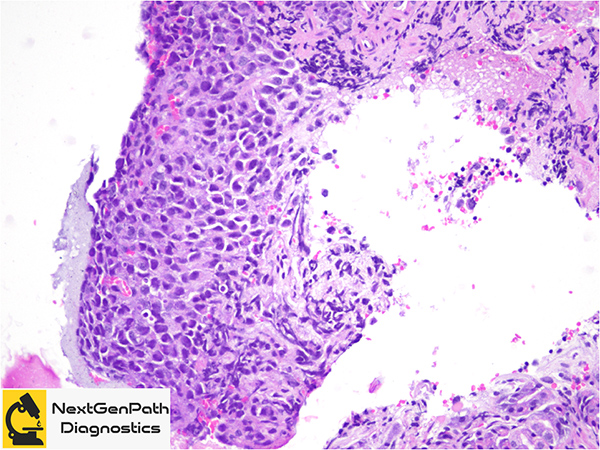
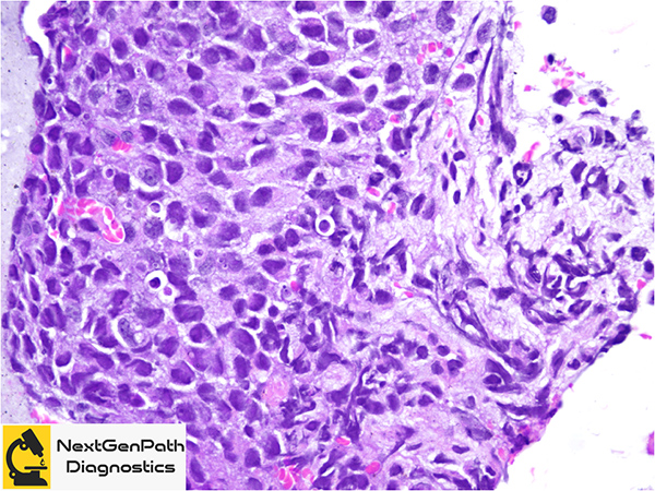
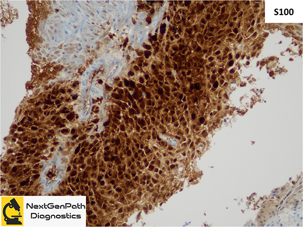
Initial evaluation should include upper endoscopy and biopsy and immunohistochemistry to make a diagnosis.
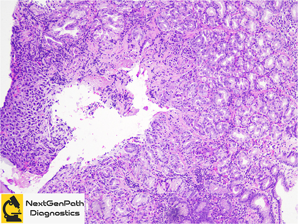
These tumors are generally located in the middle and lower esophageal lumen, are polypoid and can measure up to 5 cm.
Approximately, 70% of these tumors are non-ulcerated and pigmented, while the rest can be amelanotic and necrotic.
Although most PEM are non-metastatic at presentation, re-assessing the patient with a thorough physical exam and whole body PET-CT is necessary to rule out other primary tumors and to stage the current disease.
Given the paucity of cases, no general consensus exists regarding the optimal management and prognosis of PEM.
References
Hussein Agha Y, Parker NA, Alderson J. Case Report: Primary melanoma of the gastroesophageal junction. F1000Res. 2020 Jun 2;9:490.
Ishizaki M, Aibara Y, Furuya K. Primary malignant melanoma of the esophagogastric junction: Report of a case. Int J Surg Case Rep. 2013;4(8):700-3.