Anti-Mi-2 Autoantibody Associated DM
- Dermatomyositis (DM) is a heterogeneous IIM with a gamut of demographic, clinical, laboratory, and myopathological
features.
- The autoantibodies observed in DM are directed against various intracellular molecules, viz., Mi-2, MDA5, SAE, NXP2,
and TIF-1γ.
- Mi-2 autoantibodies were first identified in 1976, in the serum of a 60-year-old woman with features of DM (patient Mi).
- Mi-2 protein is an integral part of the nucleosome remodeling deacetylase (NuRD) complex that carries out both ATP-dependent chromatin remodeling and histone deacetylase activities.
- The characteristic presentation of anti-Mi-2 DM is proximal muscle weakness associated with skin lesions.
- Typical skin findings include Gottron papules, heliotrope rash, and shawl sign.
- Other than the skin and muscle, generally no other organ involvement is seen.
- Serum creatine kinase (CK) values are usually very high.
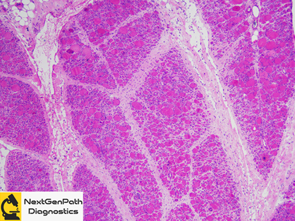
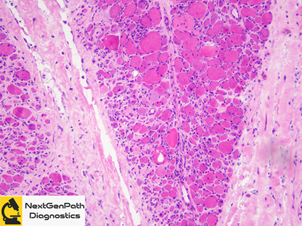
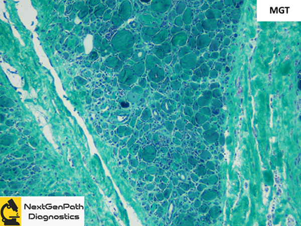
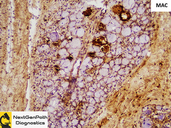
- Biopsy: Shows marked myofiber size variation, perifascicular atrophy with accompanying necrotic
myofibers and myophagocytosis . Inflammation is typically intense and diffuse comprising of lymphocytes and macrophages
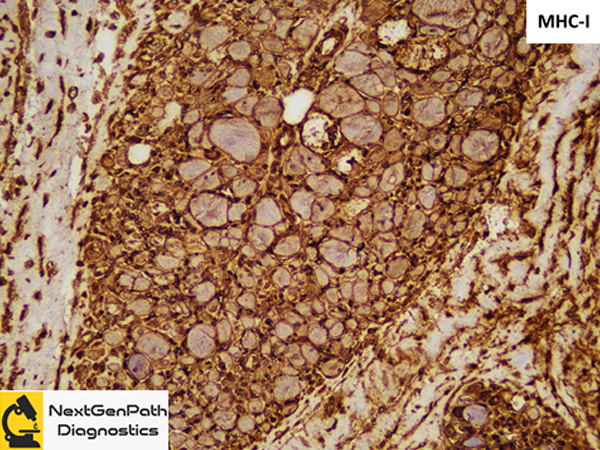
distributed in the perimysium and endomysium. MHC class I immunostaining highlights the perifascicular accentuation
gradually losing the intensity toward the center of the fascicle. Membrane attack complex (C5b-9) immunostaining
is found on the sarcolemma and less frequently on the capillaries.
- Data with respect to the association between anti Mi-2 antibodies and malignancies is controversial.
References
- Gaspar BL, Vasishta RK, Radotra BD. Myopathology: A Practical Clinico-Pathological Approach to Skeletal Muscle Biopsies. Springer Nature: Singapore, 2019.